Fetal Circulatory System: Structure, Features and Blood Flow
Before birth, a baby depends completely on the placenta for oxygen and nutrients. The lungs are not yet responsible for breathing, so blood follows a unique pathway that differs from circulation after birth. This specialized arrangement allows oxygen-rich blood from the mother to reach the baby’s organs while avoiding the lungs which are still developing.
The fetal circulatory system is designed to support growth inside the womb. It ensures that vital organs such as the brain, heart and liver receive the oxygen and nutrients needed for healthy development. Every blood vessel and temporary opening has a specific purpose during pregnancy.
Key Parts That Keep Blood Moving
Several important structures work together to maintain normal fetal circulation.
Placenta
The placenta serves as the baby’s source of oxygen and nutrients. It also removes carbon dioxide and waste products from the baby’s blood. Although the mother’s and baby’s blood stay separate, nutrients and oxygen pass through the placental membrane.
Umbilical Cord
The umbilical cord connects the baby to the placenta. It contains one umbilical vein and two umbilical arteries.
- The umbilical vein carries oxygen-rich blood to the baby.
- The umbilical arteries return oxygen-poor blood to the placenta.
These blood vessels keep the circulation process active throughout pregnancy.
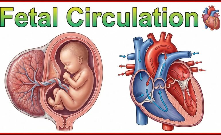
Blood Flow Pathway Before Birth
Blood follows a carefully organized route before birth.
Oxygen-rich blood enters through the umbilical vein. Most of this blood passes through the ductus venosus, which allows it to bypass much of the liver and reach the inferior vena cava quickly.
The blood then enters the right atrium of the heart. Instead of flowing to the lungs, a large portion moves through the foramen ovale, a temporary opening between the right and left atria.
From the left atrium, blood enters the left ventricle and is pumped into the aorta, delivering oxygen-rich blood to the brain and upper body.
Blood returning from the upper body enters the right ventricle. Since the lungs are not functioning, most blood passes through another temporary vessel called the ductus arteriosus, which connects the pulmonary artery to the aorta. The blood finally returns to the placenta through the umbilical arteries.
This continuous circulation supports normal fetal development.
Unique Features Before Birth
Several temporary structures make fetal circulation different from adult circulation.
Ductus Venosus
This vessel allows blood to bypass most of the liver. It speeds up the delivery of oxygen-rich blood to the heart.
Foramen Ovale
This opening allows blood to move directly from the right atrium to the left atrium without entering the lungs.
Ductus Arteriosus
This vessel connects the pulmonary artery to the aorta. It directs blood away from the lungs because breathing has not yet started.
These temporary pathways close naturally after birth as the baby’s lungs begin working.
Placental Support for Healthy Fetal Growth
The placenta performs several life-supporting functions throughout pregnancy.
It supplies oxygen, glucose, vitamins, minerals and other nutrients. At the same time it removes carbon dioxide and waste materials produced by the fetus.The placenta also produces hormones that help maintain pregnancy and support fetal growth.
Without a healthy placenta the baby cannot receive the resources needed for proper development.
Oxygen Delivery Through the Placenta
The fetus does not breathe air inside the womb. Oxygen reaches the baby through the mother’s blood.After oxygen enters the placenta, it moves into the baby’s bloodstream through the umbilical vein. The heart then distributes oxygen-rich blood throughout the body using specialized pathways.
This process continues until birth when the lungs take over oxygen exchange.
Blood Flow Changes Immediately After Birth
The biggest changes happen within minutes after delivery.When the baby takes the first breath, the lungs expand and begin exchanging oxygen.Blood starts flowing normally through the lungs instead of bypassing them.
Several temporary fetal structures close naturally:
- Foramen ovale closes.
- Ductus arteriosus closes.
- Ductus venosus closes.
- The umbilical cord no longer carries blood.
These changes create the normal circulation found in children and adults.
Supporting Growth Before Birth
Efficient circulation is essential for normal fetal growth.
A properly functioning circulatory system supports:
- Brain development
- Heart function
- Organ growth
- Muscle development
- Healthy oxygen supply
- Nutrient delivery
Any disruption in blood flow may affect fetal development and pregnancy outcomes.
Issues That May Affect Fetal Blood Flow
Some medical conditions can interfere with normal blood flow.
Examples include:
Congenital Heart Defects
Structural heart problems may alter blood circulation before or after birth.
Placental Insufficiency
Reduced placental function limits oxygen and nutrient delivery.
Umbilical Cord Problems
Compression or abnormalities in the umbilical cord can reduce blood flow.
Growth Restriction
Poor circulation may slow fetal growth and development.
Key point to know:Doctors monitor these conditions using ultrasound, Doppler studies and fetal heart monitoring during pregnancy.
Medical Monitoring During Pregnancy
Healthcare providers regularly evaluate fetal circulation throughout pregnancy.
Common monitoring methods include:
- Ultrasound examinations
- Doppler ultrasound
- Fetal echocardiography
- Prenatal screening tests
- Routine pregnancy checkups
These tests help identify circulation problems early and support timely medical care.
Supporting Fetal Growth Through Maternal Health
The baby’s circulation depends heavily on the mother’s health.
Healthy habits that support fetal development include:
- Eating a balanced diet
- Drinking enough water
- Attending regular prenatal visits
- Taking prescribed prenatal vitamins
- Avoiding smoking and alcohol
- Managing chronic medical conditions
Good maternal health helps maintain healthy blood flow throughout pregnancy.
Summary
The fetal circulatory system is a specialized network that supports life before birth by delivering oxygen and nutrients through the placenta. Temporary structures such as the ductus venosus, foramen ovale and ductus arteriosus allow blood to bypass the lungs while they develop. After birth these pathways close naturally as the lungs begin functioning. Healthy fetal circulation plays a critical role in normal growth, organ development and overall pregnancy outcomes.
Frequently Asked Questions
1. Why is the fetal circulatory system different from adult circulation?
It bypasses the lungs because the fetus receives oxygen from the placenta instead of breathing air.
2. What are the three unique structures in fetal circulation?
The ductus venosus, foramen ovale and ductus arteriosus are temporary structures that support blood flow before birth.
3. What happens to fetal circulation after birth?
The temporary openings close naturally and blood begins flowing through the lungs for oxygen exchange.
4. Why is the placenta important in fetal circulation?
The placenta provides oxygen and nutrients while removing waste products from the baby’s blood.
5. How do doctors check fetal circulation during pregnancy?
Doctors use ultrasound, Doppler ultrasound, fetal echocardiography and routine prenatal examinations to monitor blood flow.