


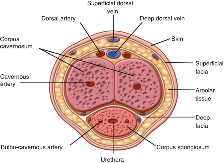
The anatomy of penis includes erectile tissues, blood vessels, nerves, muscles, connective tissues and the urethra. These structures work together for urination, erection, sexual function, and reproduction. The penis has three main regions: the root, shaft and glans.
Main Parts of the Penis
The external structure can be divided into three major parts:
- Root: The attached portion at the base of the penis.
- Body or shaft: The elongated middle portion.
- Glans: The enlarged end, commonly called the head.
Must-Know Facts:These parts connect with deeper tissues and structures inside the pelvis. The shaft contains the main erectile bodies while the glans forms the distal end of the corpus spongiosum.
The Root and Its Supporting Structures
The Root of penis is not normally visible because it lies within the pelvic and perineal region. It provides an important attachment point for the erectile bodies and associated muscles.
The root includes:
- The bulb of the penis
- The two crura
- Bulbospongiosus muscle
- Ischiocavernosus muscles
- Supporting connective tissues
Don’t Miss These Facts:The crura attach the corpora cavernosa to the pelvic structures, while the bulb forms the proximal part of the corpus spongiosum. Together these structures anchor the penis and contribute to erectile function.
The Shaft and Erectile Bodies
The shaft contains three major erectile cylinders:
- Two corpora cavernosa
- One corpus spongiosum
The corpora cavernosa lie alongside each other toward the upper or dorsal side. The corpus spongiosum runs along the lower or ventral side and surrounds the urethra.
Must-Know Facts:These tissues contain spaces that can fill with blood. During an erection increased blood flow expands these spaces and makes the penis larger and firmer.
Corpora Cavernosa: The Main Erectile Tissue
The two corpora cavernosa form much of the erectile tissue in the shaft. Each contains vascular spaces that can expand when blood enters the erectile tissue.
A tough connective-tissue covering called the tunica albuginea surrounds each corpus cavernosum. This layer provides structural support and helps maintain pressure inside the erectile chambers during an erection.The corpora cavernosa are therefore important for achieving and maintaining penile rigidity.
Corpus Spongiosum and the Urethra
The corpus spongiosum is the third erectile body. Unlike the paired corpora cavernosa, it surrounds the urethra.
Its main roles include:
- Surrounding and protecting the urethra
- Participating in erectile function
- Expanding at its end to form the glans
- Providing a pathway through which urine and semen leave the body
Key Points to Know:The corpus spongiosum does not become as rigid as the corpora cavernosa. This helps keep the urethral passage open during erection.
Glans and Urethral Opening
The glans is the enlarged end of the penis. It contains the external opening of the urethra, known as the urethral meatus.
The urethra carries:
- Urine from the bladder
- Semen during ejaculation
Important Things to Know:The urethra therefore has both urinary and reproductive functions. However urine and semen do not normally pass through the urethra at the same time. During ejaculation, physiological mechanisms help direct semen through the urethral passage.
Foreskin and Penile Surface
In an uncircumcised penis the foreskin covers the glans. It is a movable layer of tissue that can retract to expose the glans.
The Penis skin anatomy includes relatively loose and flexible skin over the shaft. This flexibility allows the skin to accommodate changes in penile size during an erection. The deeper tissues include fascial layers that provide additional structural support.Skin appearance, pigmentation and texture can naturally vary between individuals.
Internal Connective-Tissue Layers
Several connective-tissue layers support the erectile structures.
Important layers include:
- Tunica albuginea: Surrounds the corpora cavernosa and contributes to rigidity.
- Buck’s fascia: A deeper fascial layer that surrounds the erectile bodies.
- Dartos fascia: Located closer to the superficial tissues.
- Suspensory ligament: Helps support the penis from the pubic region.
These layers protect internal structures and help maintain their anatomical position.
Blood supply of penis
As part of the anatomy of penis , blood reaches the penis mainly through branches of the internal pudendal artery. The deep arteries supply the corpora cavernosa and fill the erectile spaces with blood. During erection, reduced venous outflow helps maintain firmness while veins drain blood when the erection ends.
Blood Flow and Penile Erection
An erection involves coordinated activity between the brain, nerves, blood vessels, and erectile tissue.
The basic process includes:
- Sexual stimulation activates nervous pathways.
- Signals cause the smooth muscle within penile blood vessels and erectile tissue to relax.
- Blood flow into the erectile spaces increases.
- The corpora cavernosa expands as they fill.
- Pressure inside the erectile tissue rises.
- Venous outflow becomes restricted.
- The penis becomes firm and enlarged.
The tunica albuginea helps maintain pressure within the corpora cavernosa, supporting rigidity.
Must-Know Facts:An erection can also occur without direct sexual stimulation. Spontaneous erections are a normal physiological phenomenon and can occur during sleep or at other times.
Nerve Supply of penis
The Nerve supply of penis involves both sensory and autonomic pathways.
The dorsal nerve provides important sensory information from the penis, particularly the glans and surrounding tissues. Autonomic nerves also participate in erection and ejaculation by regulating blood vessels and smooth muscle activity.
The major neural components include:
- Pudendal nerve pathways
- Dorsal penile nerve
- Pelvic autonomic nerves
- Sensory nerve endings in the glans and penile skin
Nerve signals allow the brain and spinal cord to communicate with penile tissues. This communication is essential for normal sexual response and urinary function.
Muscles of penis
The penis itself does not contain large skeletal muscles running through the shaft. However, muscles around its root contribute to penile function.
Two important muscles are:
Bulbospongiosus Muscle
The bulbospongiosus muscle surrounds part of the bulb and corpus spongiosum. It can contract during ejaculation and can also help empty residual urine or fluid from the urethra.
Ischiocavernosus Muscle
The ischiocavernosus muscles surround the crura. Their contractions can help increase pressure within the erectile bodies and reduce venous outflow during erection.
These muscles therefore support functions rather than forming the main body of the penis.
Penis Anatomy Diagram: What It Usually Shows

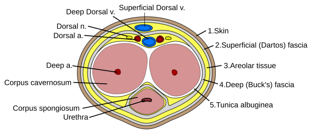
A Penis anatomy diagram typically labels the major external and internal structures so readers can understand their position.
A medically accurate diagram may identify:
- Glans
- Shaft
- Urethra
- Corpus spongiosum
- Corpora cavernosa
- Foreskin
- Root
- Bulb
- Crura
- Major blood vessels
- Supporting tissues
Key Facts You Can’t Ignore:Diagrams are particularly useful because several structures lie close together and cannot be identified from the external appearance alone.
How the Penis Supports Urination
The penis forms the final pathway through which urine leaves the body.
Urine travels:
Kidneys → ureters → bladder → urethra → external urethral opening
The urethra passes through the corpus spongiosum before opening at the tip of the glans.
The urinary function of the penis is separate from the production of urine, which occurs in the kidneys.
Penile Function in Reproduction
The penis contributes to reproduction by delivering semen into the reproductive tract during ejaculation.Sperm is produced in the testes while fluids from reproductive glands contribute to semen. The urethra then provides the final passage for semen through the penis.
Therefore the penis itself does not produce sperm. Instead it serves as an important part of the reproductive pathway.
Normal Anatomical Variation
Penile anatomy can vary considerably between individuals without indicating a medical problem.
Normal differences may involve:
- Size
- Shape
- Skin pigmentation
- Foreskin coverage
- Degree of curvature
- Vein visibility
- Glans shape
- Flaccid appearance
The penis also changes significantly between its relaxed and erect states. Loose flexible skin allows these changes to occur normally.
A person’s appearance at rest does not necessarily predict how the penis will look during erection.
Common Conditions Affecting Penile Structures
Several medical conditions can involve different parts of penile anatomy.
Examples include:
- Balanitis: Inflammation of the glans.
- Erectile dysfunction: Difficulty getting or maintaining an erection.
- Peyronie’s disease: Development of scar tissue that can cause penile curvature.
- Priapism: A prolonged erection that requires medical attention.
- Hypospadias: A congenital condition in which the urethral opening develops in an atypical location.
- Penile injury: Trauma can damage erectile tissue or other structures.
The symptoms and treatment depend on the underlying condition.
Medical Advice
A healthcare professional should evaluate symptoms such as:
- Persistent or significant penile pain
- A new lump or unusual swelling
- Blood in urine or semen
- Discharge from the urethra
- Significant changes in the skin
- Painful urination
- A sudden or severe change in penile curvature
- An erection that lasts unusually long and does not resolve
Must-Know Facts:Some penile conditions require prompt treatment, particularly prolonged erections or significant injuries.
Connection Between Structure and Function
Understanding normal anatomy makes it easier to recognize meaningful changes.
It can also help explain why several body systems influence sexual function. An erection does not depend on one structure alone. It requires coordination between:
- The nervous system
- Blood vessels
- Erectile tissue
- Connective tissue
- Pelvic muscles
- Hormonal and psychological factors
For this reason, problems affecting circulation, nerves, tissue structure or other parts of the body can sometimes affect erectile function.
Sum up
The anatomy of penis includes the root, shaft, glans, erectile tissues, urethra, blood vessels, nerves and supporting muscles. Each structure has a specific role in erection, urination and reproduction. Understanding these parts helps explain normal penile function and common medical conditions.
Frequently Asked Questions
What are the three main parts of the penis?
The three main parts are the root, body or shaft and glans.
What is the main erectile tissue?
The two corpora cavernosa form the primary erectile tissue responsible for most penile rigidity.
What does the urethra do?
The urethra carries urine and semen out of the body through the external opening at the glans.
Does the penis contain muscles?
The shaft does not contain large skeletal muscles but muscles around the root, especially the bulbospongiosus and ischiocavernosus, support erectile and ejaculatory functions.
What causes an erection?
An erection develops when nervous signals cause increased blood flow into the erectile spaces while mechanisms that restrict venous outflow help maintain rigidity.
Is penile variation normal?
Yes. Size, shape, skin appearance, curvature and foreskin coverage can naturally vary between individuals. However sudden changes, pain, swelling or other new symptoms should receive medical evaluation.
home….